What is Retina?
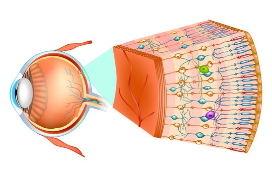
The Retina is a layer of tissue in the back of your eye that senses light and sends images to your brain. The Retina acts as a transmitter and consists of neural and blood vessel elements. It facilitates the brain to understand, and comprehend and provides a sharp, central vision needed for reading, driving, and seeing fine detail. Retinal disorders like Retinal Detachment can affect this vital tissue, thus causing problems with vision.
The Retina consists of nine inner layers interconnected with synapses and one outer layer.

What is Retinal Detachment?
Watch more videos like this on Shekar Eye Hospital YouTube Channel
Retinal Detachment is considered an ocular medical emergency caused when the 9 inner layers separate from the outer layer, due to various factors inside the eye. Retinal Detachment separates the layer of blood vessels that provides oxygen and nourishment for the cells in the retina.
Retinal Detachment is of three types:-
- Rhegmatogenous Retinal Detachment
- Tractional Retinal Detachment
- Exudative Retinal Detachment.
Rhegmatogenous Retinal Detachment:-
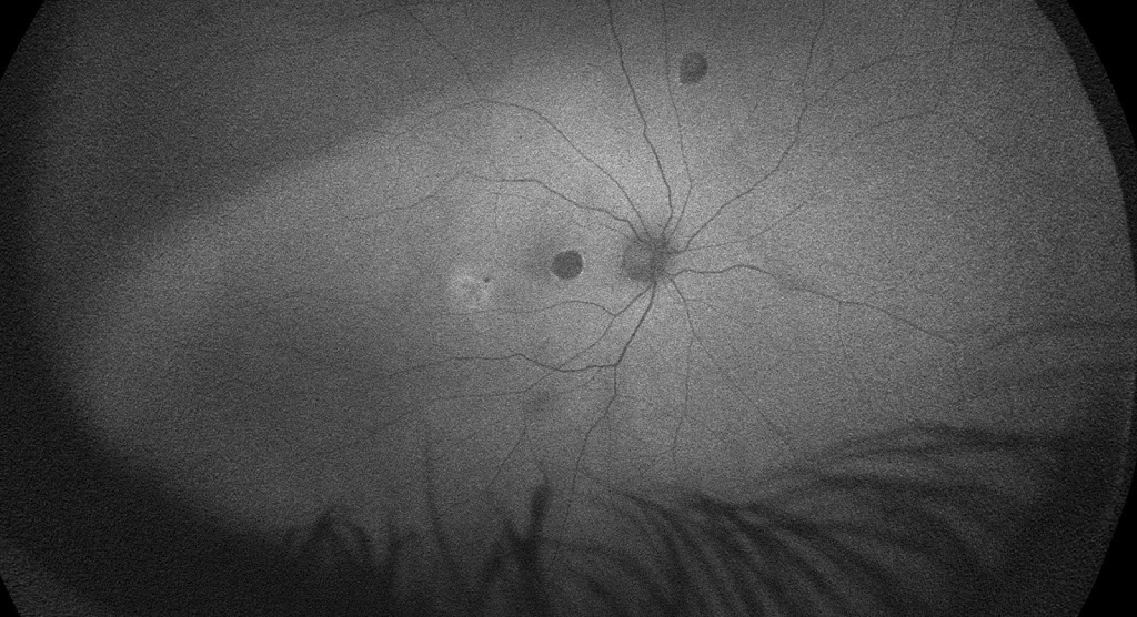
This type of Retinal Detachment is caused due to Rhegma,(Rhegma is a Greek word for break) a hole or a break in the inner nine layers of the Retina. The fluid in the eye cavity seeps through the hole, thus separating the inner layers of the retina from the outer layer, leading to a Rhegmatogenous Retinal Detachment.
Tractional Retinal Detachment:-
Tractional Retinal Detachment is most commonly caused due to diabetic and/or hypertensive retinopathy or other retinal vascular problems such as inflammation or infection of retinal blood vessels. In this type of retinal detachment, the retina is pulled due to contracting membranes which causes traction (or pull), leading to the separation of retinal layers, thus known as Tractional retinal displacement.
Exudative Retinal Detachment:-

Exudative is primarily developed due to variations in the fluid balance, secondary to various diseases in the body such as anaemia, protein deficiency, or liver damage. Since Exudative Retinal Detachment is due to other issues in the body, it is generally managed medically. It does not require any surgical treatment. The ophthalmologist will diagnose and recognize the condition and attribute it to self-healing without treating it locally.
Rhegmatogenous retinal Detachment and Tractional retinal Detachment can be treated using surgical methods.
Who are the people at risk for developing Retinal detachment ?
- Elderly : In older adults, there is vitreous degeneration and vitreous detachment. As we grow old, the homogeneous (or uniform) vitreous gel inside the eye attached to the Retina, loosens and tends to separate from it, which may cause a tear in the Retina and allows the liquified vitreous gel to seep through the layers of the Retina, causing Retinal Detachment.
- People with myopia: People with minus power of eyesight have big or oversized eyeballs, due to which there can be thinning of retinal tissue in peripheries as compared with others without any power.
- People with a family history of retinal detachment.
- People with a history of Retinal Detachment in one eye, will have the risk of developing the same in the other eye.
- People with previous eye injuries.
- People with eye diseases like uveitis, retinoschisis, or peripheral Retina thinning.
- People with previous eye surgery like cataract removal.
Symptoms of Retinal Detachment
- The sudden appearance of flashes or floaters of red in the field of vision.
- Photopsia- Light flashes visible in one or both eyes.
- Reduced side or peripheral vision
- Blurred vision

What would be the treatment modality?
Retinal Detachment is treated surgically after controlling systemic problems like diabetes, hypertension, heart or liver problems.
In cases of Rhegmatogenous RD, the retinal hole or tear has to be closed and the fluid drained so that retina gets attached. The attached retina is supported either from outside the eye in the form of a band around the eyeball or from inside the eye with the help of something called Silicon oil.

In few suitable cases of Rhegmatogenous RD, the detached retina can be settled using a gas bubble that will absorb by itself in 15-20 days.
For Tractional RD, the surgery is always from inside the eyeball, involves Vitrectomy(removing the gel of the eye) followed by careful dissection of the membranes that pull the retina causing detachment and finally supporting the attached retina with Silicon oil.
Retinal detachment surgeries have been around from 1960 to 1970, and on the current day, there is nearly 95%-98% success rate in the attachment of Retina restoring its structure.
When to see a doctor?
Retinal Detachment can be treated and managed well with proper diagnosis and timely intervention. Consult a Retina specialist immediately when you experience any of the signs or symptoms of Retinal Detachment.


