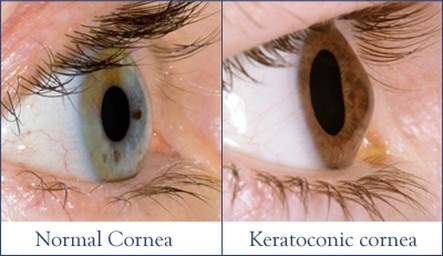
Keratoconus, often abbreviated to “KC”, is a non-inflammatory eye condition in which the normally round dome-shaped cornea progressively thins causing a cone-like bulge to develop. This results in significant visual impairment.
Keratoconus is generally first diagnosed in young people at puberty or in their late teen’s. It has no known significant geographic, cultural or social pattern.
Keratoconus Symptoms – What Happens?
Corneal Bulging
The cornea is the clear window of the eye and is responsible for refracting most of the light coming into the eye. It is the front part of the eye. This part is clear, transparent and spherical in shape in the centre. This curvature is important for the light rays to be focused on to the sensitive parts of the retina to form clear image. Therefore, abnormalities of the cornea severely affect the way we see the world making simple tasks, like driving, watching TV or reading a book difficult. When this shape of cornea is altered leading to cone like protrusion in the front of cornea condition is termed as Keratoconus.
In its earliest stages, keratoconus causes slight blurring and distortion of vision and increased sensitivity to light. These symptoms usually first appear in the late teens and early twenties. Keratoconus may progress for 10-20 years and then slow or stabilize. Each eye may be affected differently.
Cornea is made up of tiny fibers arranged in specific manner called as collagen fibers. When this collagen fibers are weak and cannot hold cornea in specific shape, it may lead to weakness of the cornea and change the shape of cornea. This change may progress making cornea cone shape. This is attributed to decrease in anti oxidants level, making corneal collagen weak.
What Causes Keratoconus?
Keratoconus is a multifactorial disease and may run in families as well. This may also be seen in some of the systemic diseases mostly with connective tissue disorders. Condition is seen as part of many genetic syndromes also. It can also be result of chronic eye problems in which there is intense rubbing leading to change in curvature of the disease.
Most commonly condition starts in the young age , either childhood or teenage, may occur in any age group also. Change in the shape may not be sudden onset, but usually progresses slowly over months or years. Condition usually affects both the eyes and progression is faster in younger age group.
Patients may have blurring of vision due to change in the curvature of the corneal surface leading to astigmatism, which is irregular astigmatism. Patients may also develop nearsightedness due to expansion of the cornea. Sudden change in the refractive power may be present.
Some patients may also have double vision, haloes, difficulty in driving. In cases of allergic conjunctivitis, intense itching, foreign body sensation, redness may be present, that can lead to intense rubbing of eyes and change in curvature of the disease. In severe condition, cornea may bulge with thinning leading to rupture and the forming scar.
Keratoconus – Diagnosis
Diagnosis of the condition is based on various clinical examination and various tests. Slit lamp examination can pick various signs present in the disease. Retinoscopy is done to determine the refractive error , which may also show signs of keratoconus. Other tests include keratometry done to look for curvature of the cornea in vertical and horizontal axis. Imaging techniques such as topography and PENTACAM can give information about curvature at every point of cornea, accurate astigmatism, corneal thickness. All these tests are required for the diagnosis, treatment and prognosis of the condition.
Keratoconus Treatment: What Can Be Done About it?
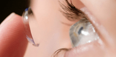
Various treatment options are available for the disease no based on the severity of the disease. Early keratoconus can be treated with spectacles and rigid contact lenses to correct the astigmatism and give clear vision. Various types of contact lenses are available for the treatment of keratoconus. But disadvantage of rigid contact lenses is that, in severe cases optimal fit may not be achieve and become difficult to wear.
In the early stages, eyeglasses or soft contact lenses may be used to correct the mild shortsightedness and astigmatism caused in the early stages of keratoconus.
As the disorder progresses and the cornea continues to thin and change shape, rigid gas permeable (RGP) contact lenses are generally prescribed to correct vision more adequately.
Newer lenses like Rosek, Kerasoft and Scleral contact lenses are available for better vision and comfort. The contact lenses must be carefully fitted, and frequent checkups and lens changes may be needed to achieve and maintain good vision. Intacs (Intracorneal rings) are sometimes used to improve contact lens fit.

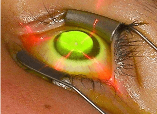
COLLAGEN CROSS LINKING is the procedure done to halt the progression of the disease. It is based on the collagen cross linking with ultra violet A and riboflavin. This causes change in biomechanics of the corneal collagen and hence strengthening the cornea. However, this procedure may need to be repeated if necessary.
INTACS are the acrylic rings inserted in the corneal layers. They can improve vision and also can be combined with collagen cross-linking.
KERATOPLASTY or corneal transplant is the surgical treatment that is reserved for advanced cases of keratoconus. The central portion of the cornea is replaced with a donor cornea. Surgery although gives good results, is associated with risk and complications.

Keratoconus is the corneal condition due to change in the shape of the cornea, usually seen in young age, associated with many symptoms that can be treated with contact lenses and various surgical procedure. Topography and imaging techniques is needed in cases of astigmatism to detect this condition early and give necessary treatment.


