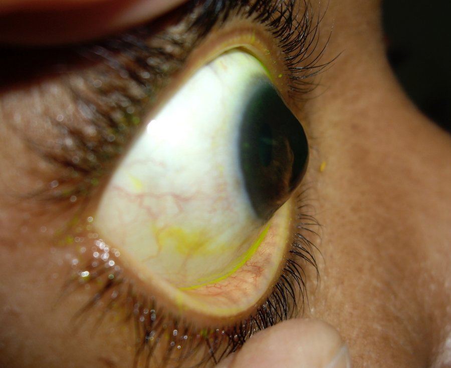
What is keratoconus?
Do you have any irregularities in the corneal surface, or does your cornea bulge out? If yes, you might be experiencing symptoms of keratoconus, an eye disease. As we all know, the eye is a complex organ made up of several layers.
Keratoconus is a progressive disease of the eye characterized by a thinning of the cornea, the clear outer lens or “windshield” of the eye. This can transform the cornea from a symmetrical dome to an asymmetric cone. Keratoconus means conical cornea in Greek.
The main function of your cornea is to refract light into your pupil. Therefore, light passing through your asymmetrical cornea can lead to distortion and blurriness in your vision.
The tiny fibers of protein in the eye called collagen help hold the cornea in place. However, the low level of protective antioxidants weakens these fibers, as a result of which they can’t keep their round shape and bulge outward like a cone.
It most often develops during your teenage or young adulthood. It tends to get progressively worse for 10 to 20 years before stabilizing and tends to be more aggressive in children than adults.
Causes of Keratoconus
The definitive cause of keratoconus is unknown, though it is believed that the predisposition to develop the disease is present at birth. The possible causes include:
Family history:
For some people, keratoconus may carry genes that make them predisposed to its development if exposed to certain environmental factors.
Underlying disorders:
Keratoconus sometimes may occur in the presence of certain underlying disorders such as Down syndrome, sleep apnea, asthma, some connective tissue disorders including Marfan syndrome and brittle cornea syndrome, and Leber congenital amaurosis. Note that a direct cause and effect hasn’t been established.
Inflammation:
In some cases, inflammation such as allergies, asthma, conjunctivitis, eczema, hay fever or atopic eye disease can damage the tissue of the cornea.
Eye rubbing:
Rubbing eyes harder can result in the progression of keratoconus for people who already have it. This is because rubbing eyes hard over time can break down the cornea.

Symptoms of keratoconus
The most important sign of keratoconus is a thinning of your cornea that disrupts its natural dome shape. Although, initially, it’s common not to have any symptoms; as the condition progresses, it shows the following symptoms:
- Double vision when looking with just one eye
- Objects both near and far that look blurry
- A persistent desire to rub your eyes
- Frequent changes in spectacle power
- Distorted vision or Shadow vision
- Nearsightedness (difficulty seeing far away)
- High cylindrical power for the spectacle.
- Blurry vision that makes it hard to drive
- Halos in your vision
- Eye strain
- Irritation

Symptoms may start in one eye, but they eventually affect both eyes in most cases.
Diagnosis
An ophthalmologist or cornea specialist will give you a thorough eye examination and check your medical and family history for the diagnosis of keratoconus. During eye examination, your eye doctor will examine shape and thickness of the cornea. Visual acuity is checked, sometimes to check the best corrected acuity contact lenses will be required.
Corneal topography is a computer-assisted diagnostic tool that creates a three-dimensional map of the surface curvature of the cornea. It allows your doctor to examine changes to your eye that aren’t otherwise visible.
Management of Keratoconus
Keratoconus treatment focuses on the correction of vision and depends on the severity of the condition and how fast it’s progressing.
In the initial stages, spectacles and soft contact lenses can be used to correct the vision. When your symptoms are mild, your vision can be corrected with eyeglasses. However, as keratoconus worsens, vision may no longer be correctable with a soft contact lens; for such patients, doctors recommend a special type of hard contact lens.
Different types of contact lenses include Rigid gas permeable contact lenses, Hybrid lenses, Scleral lenses etc. A contact lens can also be used in the case of advanced cases along with other lines of treatment.
The earlier the onset of keratoconus, the faster the progression. The frequent change of spectacle power indicates the progression of keratoconus. Progressive keratoconus can be treated by a procedure called Corneal Collagen Cross-Linking with Riboflavin (C3R).
This is a minimally invasive procedure in which the doctor uses Vitamin B eyedrops in your eye. The eye is then exposed to ultraviolet light for up to 30 minutes to strengthen the bonds between the cornea’s collagen fibres and surrounding proteins to prevent further thinning or bending.
In advanced cases of keratoconus where vision can no longer be corrected with the above treatments, surgeries like Intracorneal ring segment, Corneal transplant, or keratoplasty are recommended. In this case, the doctor will remove the centre of your cornea, replace it with one from a donor, and stitch the new one into place. You may need contact lenses afterwards.
It’s very important to get the treatment as early as possible because once the keratoconus progresses, it deteriorates the vision. It’s difficult to bring back the vision with any treatment once it deteriorates. The earlier the procedure is done, the better the vision. Keratoconus is a treatable condition but make sure that if you or any of your close family members have potential keratoconus symptoms, it’s essential to visit your eye doctor for a proper examination. The earlier the diagnosis and treatment for keratoconus, the better the prognosis. Thanks for reading.


